Brain Aneurysm Diagnosis
Diagnostic assessments typically involve:
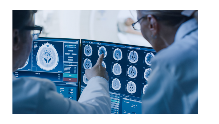
Computerized tomography (CT). A CT scan, a specialized X-ray exam, is frequently the first test used to verify if there is bleeding in the brain. The exam produces images which look like 2-D “slices” of the brain.
Patients may possibly also receive an injection of a dye to make easier the blood flow visualization in the brain which may perhaps indicate the presence of an aneurysm. This variant of the exam is called so CT angiography.
Cerebrospinal fluid test. When a subarachnoid hemorrhage has occurred, there will presumably be red blood cells in the fluid surrounding the brain and spine (cerebrospinal fluid). The doctor will order a test of the cerebrospinal fluid if there are symptoms of a ruptured aneurysm although a CT scan has not exhibited proof of bleeding.
This procedure is called a lumbar puncture (spinal tap), and its aim is to draw cerebrospinal fluid from the back.

Magnetic resonance imaging (MRI). This technique uses a magnetic field and radio waves to generate complete images of the brain, 2-D slices and / or 3-D images.
MRI angiography is a kind of MRI that assesses the arteries in detail and may detect the presence of an aneurysm.
Cerebral angiogram. The doctor inserts a thin catheter into a large artery and threads it past the heart to the arteries in the brain. A special dye infused into the catheter goes to arteries throughout the brain. Then, a sequence of X-ray images can reveal details about these arteries and detect an aneurysm.
This exam is the most invasive one and it is usually used when other diagnostic assessments do not deliver enough information.
Brain Aneurysm Treatment
Nowadays, the treatment for brain aneurysms is more promising than it used to be.
Innovation and clinical evidence have developed into more effective and less- invasive treatment options for patients.
Clinicians take into consideration several factors when deciding which treatment option is best for a specific patient. Some of these factors may include
![]() The size, location, and overall aspects of the aneurysm
The size, location, and overall aspects of the aneurysm
![]() Patient age and general health
Patient age and general health
![]() Family history of ruptured aneurysm
Family history of ruptured aneurysm
![]() Congenital conditions that boost the risk of a ruptured aneurysm patient age, size, and location of the aneurysm.
Congenital conditions that boost the risk of a ruptured aneurysm patient age, size, and location of the aneurysm.
Possible treatment options:
Open surgery (clipping)
It is a surgical method where the surgeon reveals the aneurysm with a craniotomy and places a metal clip across the base of the aneurysm with the purpose of blood cannot enter it.
Endovascular therapy (coils, stents, flow diversion device, intrasaccular devices, etc.)
This option is a less invasive procedure than the previous one, where the surgeon inserts a catheter into an artery, and threads it through the body to the aneurysm.
No treatment: observation, controlling risk factors and reiterating imaging exams.
Typically, the primary goal of treatment is to prevent the aneurysm from bleeding or re-bleeding. Treatment does not frequently get better symptoms except when nerves are being pressed by the aneurysm.
Lifestyle Modifications to Reduce the Risk
When having an unruptured brain aneurysm, it is important to decrease the risk of its rupture by making some daily life shifts which include adopting healthy habits, as diet, and exercise routines.
Reference Links:
https://bafound.org/treatment/https://bafound.org/about-brain-aneurysms/brain-aneurysm-basics
https://www.mayoclinic.org/diseases-conditions/brain-aneurysm/diagnosis-treatment/drc-20361595
- Neurologic disorders (Chapter 3). In: Professional Guide to Diseases, 9th edn. Lippincott Williams & Wilkins, 2009.
